Animal models to study metastasis
Orthotopic mouse models
Orthotopic tumour models are considered more clinically relevant and better predictive models of drug efficacy than standard subcutaneous models. Since tumour cells are implanted directly into the organ of origin, these tumours reflect the original situation (such as e.g. microenvironment) much better than subcutaneous xenograft tumour models. The combination of Luciferase gene transfected cancer cells together with orthotopic implantation of these cells allows non-invasive visualization and quantification of primary tumour growth as well as metastases. Models available at LECR are colorectal (caecal injection) and breast (mammary fat pad), intraperitoneal and intracardiac models.
PDX mouse models
Patient-derived xenografts (PDXs) are prepared by direct engraftment of patient-derived tumour fragments or cell suspension orthotopically or ectopically (in general subcutaneous) into immunocompromised mice. PDX faithfully recapitulates human tumour biology.
CAM models
The chicken Chorio-Allantoic Membrane (CAM) model, the chick embryo is surrounded by the chorioallantoic membrane (CAM), a highly vascularized extra-embryonic membrane that can be used to graft human cells. When grafted on CAM, tumour cells are capable of stimulating the formation of new blood vessels, gaining their blood supply. The chick embryo is naturally immunocompetent, thus easily allowing mammal cell xenografts. The procedures are relatively simple, involving short experimental times and low costs. The use of this alternative animal model fits in the "3Rs policy".
Video derived from JOVE
Luminescent monitoring in vitro/in vivo
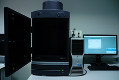
The IVIS Lumina II system includes a sensitive deep-cooled CCD camera, a light-tight imaging chamber, gas anesthesia, a temperature-controlled stage, and image analysis software. This system enables non-invasive visualization and quantification of fluorescent or bioluminescent signals a living animal. It can also be used to visualize ex vivo and in vitro samples. For in vitro luminescent analysis of well plates, if uniformly distributed, our plate reader (ParadigmTM) might be better suited.
