Additional measurement equipment
Besides the other mentioned research equipment, which are located and used by the department, also some additional measurement equipment are used frequently, but are not located in our department.
MRI
Our Department amplifies an existing collaboration with the Radiology Department (Ghent University Hospital), a major regional hospital, where we have access to a 3T high-field TIM TRIO MRI-scanner (Siemens, Erlangen). The MRI data acquisition is performed in collaboration with Prof. E. Achten, Head of the Department of Radiology at Ghent University Hospital. There is also a partnership between our research group and the Ghent Institute for Functional and Metabolic Imaging (GIfMI) on one hand, and the Multidisciplinary Research Platform (MRP) of the Institute for Neuroscience on the other hand, which provide exceptional platforms for integration of cognitive neuroscience and neuromodulation using combined resources of Ghent University and Ghent University Hospital.
Besides helping the understanding of pathological mechanisms in clinical groups, including Autism Spectrum Disorders, Traumatic Brain Injury, and Developmental Coordination Disorder, we use brain metrics as complementary biomarker to monitor training effects by using state-of-the art medical imaging techniques.
Specifically, we administer five different types of specialized MRI scans to characterize the connectome, or the ‘fingerprint’ of an individual’s brain connectivity.
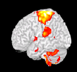
- Task-based functional MRI (T-fMRI) is used for non-invasive measurements of haemodynamic processes within the brain during the performance of a motor task.
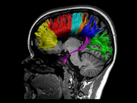
- Diffusion tensor imaging (DTI) is used for anatomical investigation of the integrity of anatomical connections by providing a quantitative assessment of the brain’s white matter microstructure.
- A magnetization transfer image (MTI) is acquired to obtain a more dedicated measure of myelin integrity.
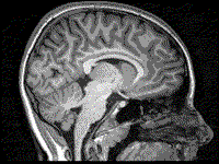
- a high-resolution T1-weighted MRI scan is administered to quantify cortical thickness and cortical surface area.
- resting state fMRI (R-fMRI) is used to evaluate specific patterns of synchronous activity that occur when a subject is not performing an explicit task.
Dexa scan
Dual-energy X-ray absorptiometry (DEXA) is a two-dimensional imaging method, which is used to evaluate bone mineral density and total body composition (lean, fat and bone mineral mass).


